
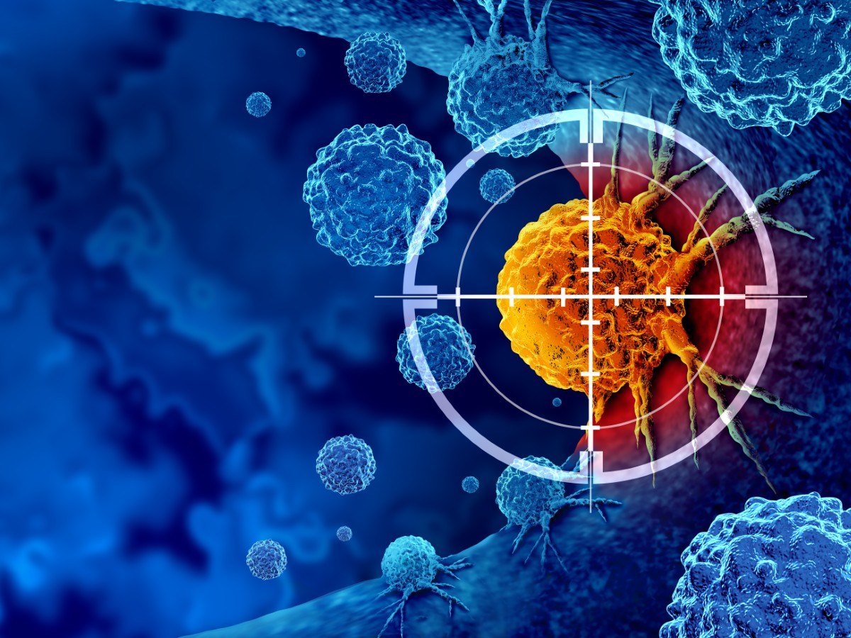
Researchers from Fudan University, Sun Yat-sen University, and the Chinese Academy of Sciences in Shanghai have discovered a mechanism by which cancer cells self-destruct when subjected to prolonged compression. Utilizing three-dimensional models, the team revealed how cancer cells respond to mechanical confinement. Their findings show that when trapped in tight spaces, these cells experience chronic stress, altering their metastatic behavior and leading to cell death. The study was published in the journal Developmental Cell on October 14.
What The Study Found
The researchers focused on cancer cells undergoing a specific developmental stage known as the mesenchymal-to-amoeboid transition (MAT). This rapid process enables cells to adapt to narrow spaces without genetic transcription. According to Liming Sun and Yan-Jun Liu, squeezing through extremely narrow spaces presents a significant challenge for cancer cells during metastasis. To study this, the researchers developed a system that gently compressed cells into thin spaces, mimicking the stressful conditions they encounter while evading the primary tumor and invading nearby tissues.
Under these conditions, researchers observed the transformation of cancer cells into fast-moving, amoeba-like states. Surprisingly, prolonged confinement caused these cells to eventually halt movement and activate a cell death program. As pressure increased, the environment was perceived as dangerous by the cells, which released signals triggering apoptosis. This continuous confinement imposed lasting stress, activating MLKL, which initiates cell death.
Before dying, the cells lost their polarized shape and mobility, ceasing invasive behavior. But this process also removed MLKL-high cells, leaving tumors enriched with MLKL-low, necroptosis-resistant clones that exhibit more aggression. This mechanism was tested on various cancer cell types, displaying similar responses. Sun and Liu noted that this might be a broader principle of tumor cell response to prolonged physical stress.
However, they warned that the response’s extent might vary based on different cancer characteristics, necessitating further studies.
What The Findings Mean
The researchers told Newsweek that prolonged mechanical stress within tumors acts as a natural barrier against cancer cell spread. It eliminates highly invasive cells and promotes immune tumor recognition. MLKL expression may serve as a biomarker for tumor progression and metastatic potential since higher MLKL expression made cells more susceptible to mechanical checkpoints.
Over time, selective pressure favors expansion of escape clones with increased migratory and invasive potential. Measuring MLKL levels can help assess tumor evolution during disease progression.
These insights could lead to new therapeutic strategies. One option involves pharmacologically activating MLKL to enhance MLKL-dependent necroptosis, potentially increasing the removal of cancer cells under prolonged compression. The study proposes combining approaches to restore or activate MLKL with conventional therapies, potentially strengthening the body’s defenses against metastasis.
“Therapies should not only target biochemical pathways but also exploit the unique mechanical environment of solid tumors,” Sun and Liu emphasized.
Such combined therapies may eventually support the innate defenses against metastasis.
The study was conducted by Zhang W, Wang Y, Zuo C et al (2026), titled “Long-term confinement triggers RIPK3-independent necroptosis in amoeboid cancer cells to shape tumor evolution,” published in Developmental Cell.